- What we are made of?
- Cells.
Due to the advent of microscope, we are able to see basic building block of organisms, cell. And, the improvement of microscopy over years made it possible to see cell in much more details now. So, the microscope is a tool for biologists, similar to the telescope for astrophysicists.
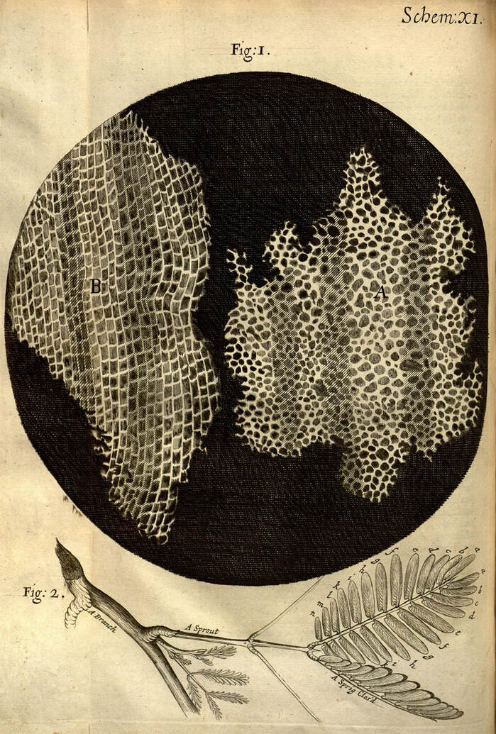
Here is the Robert Hooke's first observation of cell and drawings of the cellular structure of cork and a sprig of sensitive plant from Micrographia (1665).
When we take a look at cells from multicellular organisms like us under the microscope, we observe many many cells together.
- From where all these cells are coming?
- From another cell.
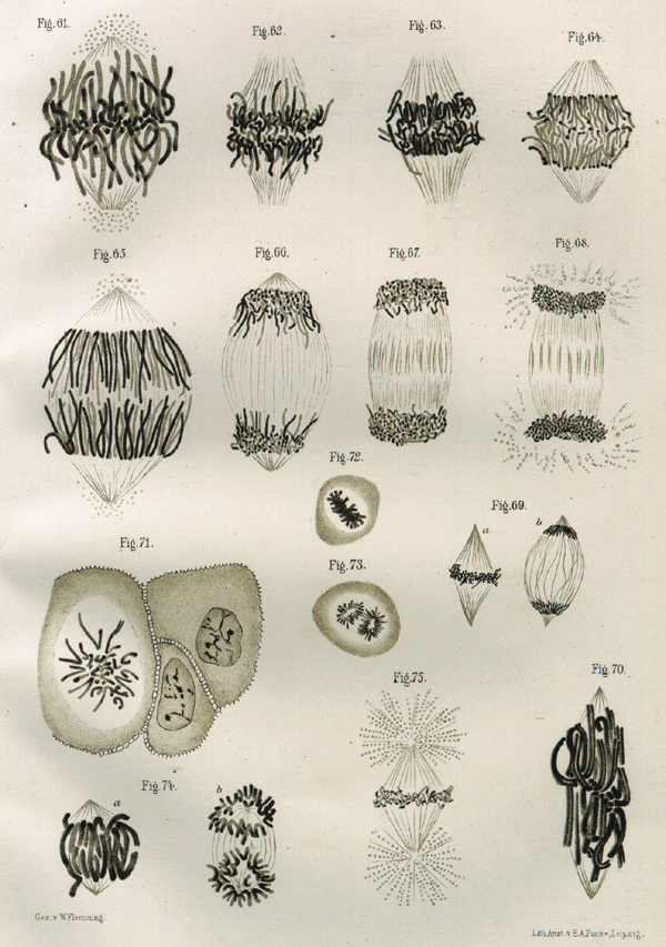
Really? How? One new cell is coming from another cell? One of the major concepts from the cell theory was: all cells are coming from another cell. Again, the favorite tool for biologists, microscope, came to rescue. Walther Fleming first observed under the microscope that a cell is going through division and producing two new cells. The source cell is known as mother cell and the newly produced cells are called daughter cells.
He used the by product of coal tar, known as aniline dye, to visualize threadlike chromosome during the cell division. Here is Walther Fleming's visualization of mitosis in 1873.
Cell division answers the question about the origin of each cells. But, we have many different cell types with different functions. If a mother cell divides and produce two daughter cells with similar function, then all the existing cells should have same function. In reality, it is not the case.
- How do we get new cell type?
- Asymmetric cell division.
Asymmetric cell division follows all the basic steps of mitosis, except the mother cell produces two daughter cells with different functions. We can distinguish two daughter cells with different function immediately either by looking at their size and shape or knowing the cell type specific genetic factors.
In 1905, Conklin first observed a "yellow-ish cytoplasmic substance" deposited in the mother cell. After the cell division process, one daughter cell inherit "yellow-ish cytoplasmic substance", but the other daughter cell doesn't! Interestingly, the daughter cell with the yellow cytoplasm develop into muscle cell.
During the asymmetric cell division or distribution of cell fate determinants, cell needs to know the direction. It is exactly the same way we use Google map on our phone. The Google map tells us which way we should go. In cell, some polarly localized proteins provide direction.
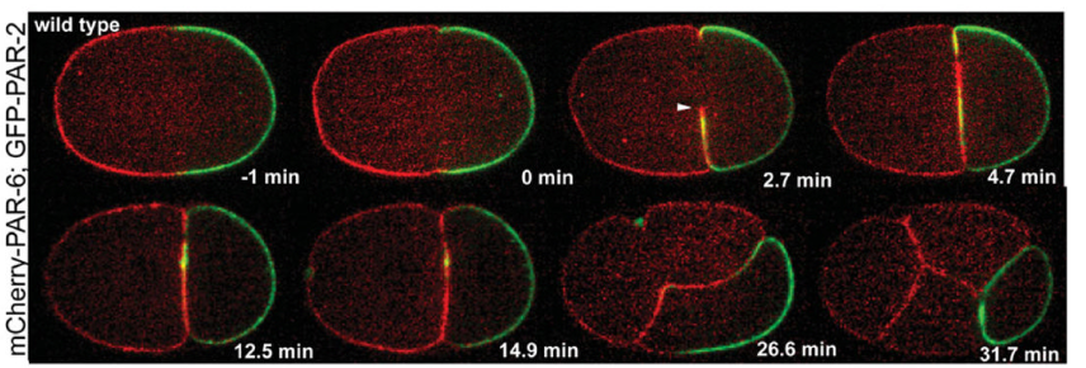
In 1988, Kemphues et al. discovered the first polarly localized protein, PAR. Here is an example of polarly localized PAR proteins.
In 1988, Kemphues et al. discovered the first polarly localized protein, PAR. Here is an example of polarly localized PAR proteins.
When we search for a direction, we have intention to use that directional information to move towards it. Similarly, these polarly localized proteins direct cellular organelle, such as nucleus. It is an ideal scenario for asymmetric cell division, where nucleus needs to move away from the center of the cell.
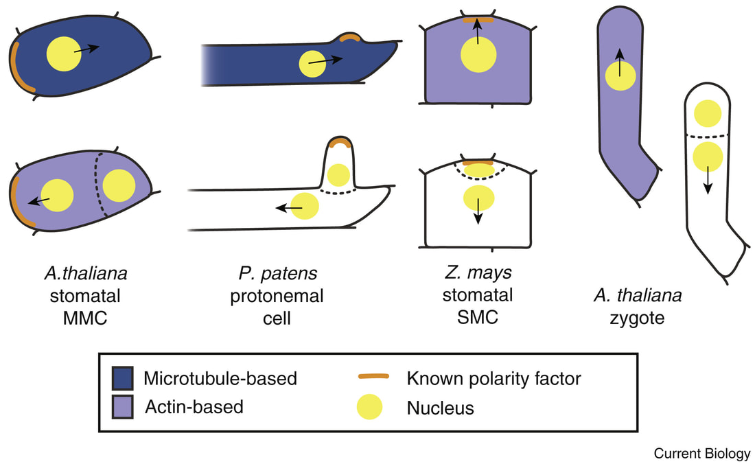
This directional nuclear movement is facilitated by the usual suspect, cytoskeleton. In cellular context, cytoskeletons act as train track for nucleus movement. Plant cell biologists took advantage of many elegant cell types to combine cell polarity, cytoskeleton-dependent nuclear movement, and asymmetric cell division.
This directional nuclear movement is facilitated by the usual suspect, cytoskeleton. In cellular context, cytoskeletons act as train track for nucleus movement. Plant cell biologists took advantage of many elegant cell types to combine cell polarity, cytoskeleton-dependent nuclear movement, and asymmetric cell division.
The next interesting question is: Who is the master regulator in this cellular process? Nucleus decides the future cell division site? Or nucleus follows the instructions to position towards the already decided future cell division site?
Classic experiments utilized drug treatment (inhibiting cellular cytoskeleton track) or external cues (centrifugation, light) tried to displace nucleus and found that the future cell division happens according to the misplaced nuclear position. But, these conditions, we not only displace nucleus, but also hampers the overall cellular activities.
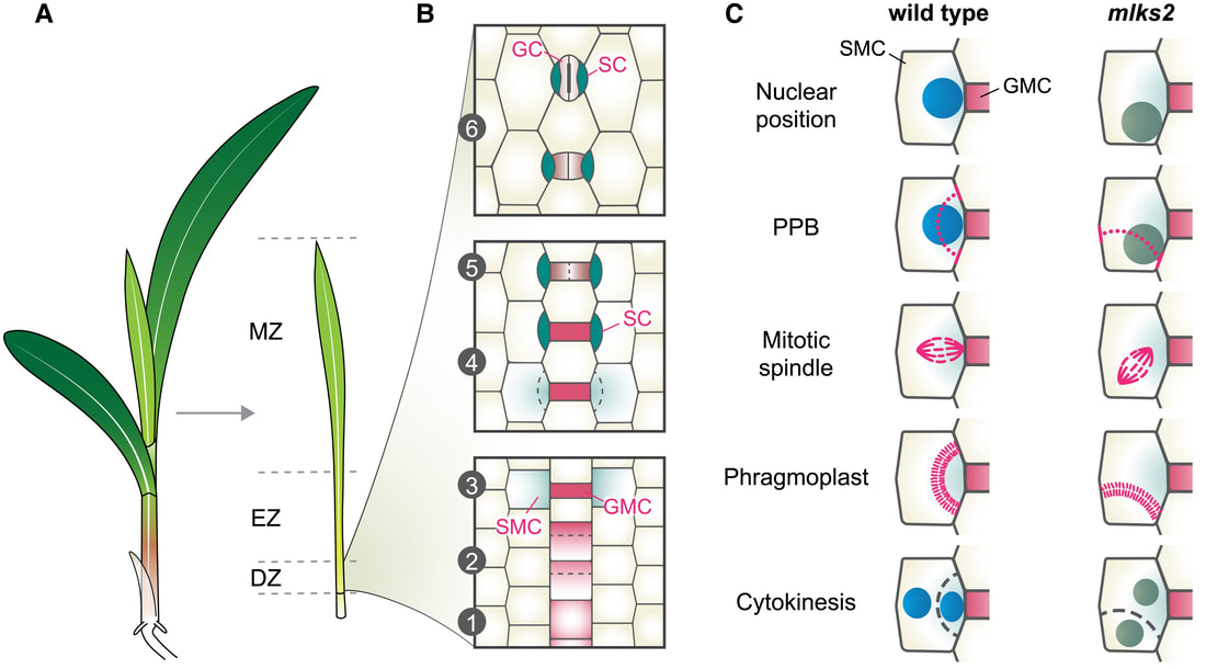
In a recent study, Arif took advantage of the nucleus envelope proteins, which connect nucleus and cytoskeleton for their movement. A mutation in the nuclear envelope protein disturb the link between nucleus and cytoskeleton, without disturbing other cellular processes. Using long-term live cell imaging, Arif discovered that when nucleus fails to go to the destination, cell decides to divide according to the misplaced nuclear position. In the following figure, Janlo Robil summarizes this exciting discovery.
